
Age-related color loss shows up most on the blue-yellow axis, and most subtle changes go unnoticed

Fig. 1: A scene rich in blue and yellow tones, the axis where age-related color loss shows up most.
Most of the conversation about color-enhancing lenses is framed around athletes and outdoor enthusiasts: sharper edges on a mountain bike trail, faster target discrimination on the range. That framing leaves out a very large group. Color vision changes with age, and most subtle changes go unnoticed unless someone is tested. When the changes do show up, they are mostly along one axis, blue-yellow.
The lens yellows gradually, and blocks more blue over time
The eye’s natural lens yellows slowly over a lifetime, and as it does it absorbs more short-wavelength, blue light and scatters more light overall. In Boettner and Wolter’s 1962 measurements of nine freshly enucleated eyes, aged 4 weeks to 75 years, the lens of a young eye reached 90 percent total transmittance by about 450 nanometers, while a 63-year-old lens did not reach 90 percent until about 540 nanometers. Older lenses also scattered more light. Because the study measured one eye per age, it shows the trend, not a population average.
It's not just the lens
Lens yellowing is only one contributor. Researchers at the Smith-Kettlewell Eye Research Institute list several factors that may affect color vision with age, blue-yellow recognition especially: a smaller pupil that lets in less light, a yellowing lens, rising rates of eye disease, and changes in the sensitivity of the visual pathways. On that last point, one study of the pathway that carries signals from the eye’s blue-sensitive cones, known as S-cones, found that the part handling decreases in S-cone stimulation responds more slowly in older observers, while the part handling increases stays stable. Retinal responses to S-cone stimulation also change with age in eyes whose natural lens has been replaced, though responses from the other cone types change too. A study of peripheral resolution found that both blue-yellow and black-and-white resolution held flat until about the fifth decade of life and then fell by roughly 14 percent per decade, and that lens density did not explain the decline in older subjects.
Researchers still debate how selective all of this is. A 2016 review argues that S-cone pathway losses are not unique to those cones and that the visual system compensates enough to keep color appearance fairly stable. A 2013 study of 185 adults likewise found that supra-threshold color perception was largely unaffected by age, even as the ability to tell apart small color differences declined.
Why blue and yellow stand out
Human color vision runs on three cone types, tuned to short, medium, and long wavelengths, compared against each other through two main channels: a red-green channel built from medium and long cones, and a blue-yellow channel that depends on the S-cones. S-cones make up less than 10 percent of the cones in the retina to begin with. In the 2014 study described below, roughly three-quarters of the people who failed a color-vision test showed blue-yellow errors, and its lead author noted that the majority of color defects among older people are of the blue-yellow type. Lens yellowing, which absorbs short wavelengths, is one plausible reason that axis is hit hard, though as above it is not the whole story.
A slow decline most people never notice
Clinical abnormalities are uncommon before 70, but measured color sensitivity starts to decline much earlier. A review of color-threshold measurements in several hundred observers aged 4 to 90 found that sensitivity was best around age 20 and then declined gradually and almost linearly, by roughly 1 percent per year for red-green and 1.6 percent per year for blue-yellow. The reviewers note that people typically remain largely unaware of this normal change because the visual system adapts, and they suggest the loss of nerve fibers from the retina may account for part of it, with reduced short-wavelength light reaching the retina as the most obvious extra factor for blue-yellow.
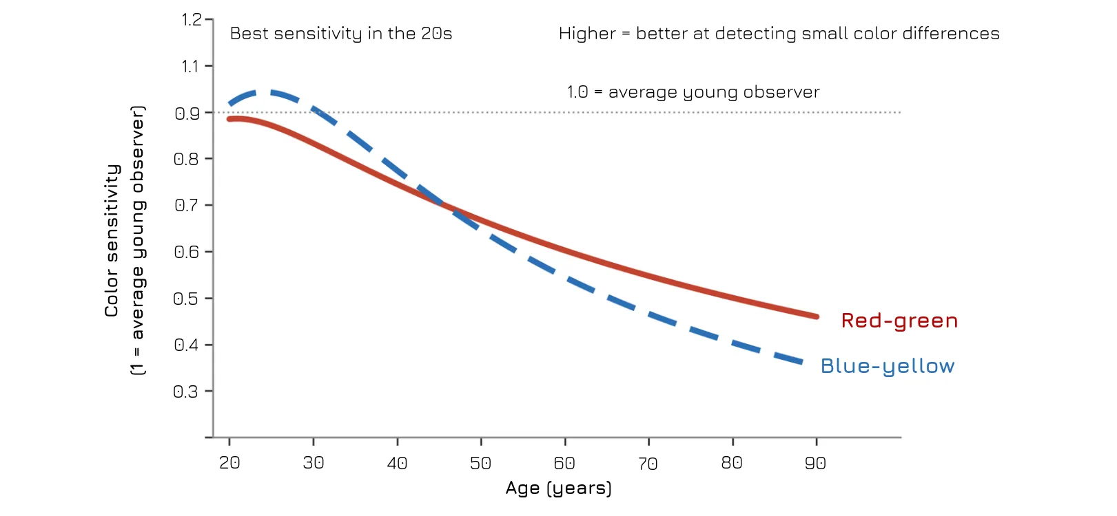
Fig. 2: Average color sensitivity declines with age, faster for blue-yellow than red-green.
How common it actually is
A study published in Optometry and Vision Science in 2014 tested a random sample of 865 adults aged 58 to 102, excluding anyone with inherited color deficiency, and found that abnormal color vision was uncommon before age 70 and then rose quickly. About 45 percent of people in their mid-70s had an abnormality, up to 50 percent of those 85 and older, and nearly two-thirds of those in their mid-90s. Overall, 40 percent failed at least one of the two color-vision tests used and about 20 percent failed both, including the easier test designed to catch defects severe enough to affect everyday tasks. The sample came from one Northern California county, so the rates are best read as a snapshot, not a global figure. Most subtle abnormalities, the authors suggest, are likely to go unnoticed.
Color vision problems are uncommon before 70. After that, the rate climbs quickly, reaching half or more of people in the oldest groups studied.
Where this leaves Colorboost
Age-related color loss is a shrinking margin for telling similar colors apart. CRF puts a number on a related property of a lens: how many meaningfully different colors it lets a wearer resolve, compared with a clear lens. CAF tracks how closely those colors stay to natural, which is worth watching when the margin is already narrowing. Colorboost’s published reference figures are a CRF of 135% and a CAF of 99% against a clear lens, with a 35% larger color volume. This article does not present data from older observers, and CRF is a general measure, not specific to the blue-yellow axis. We aren’t aware of a published study testing a color-enhancing lens on an older population, so whether it helps this group is an open question. CRF and CAF give that question numbers to be tested against.
If the blue-yellow axis is where age-related color loss shows up most, and most subtle changes go unnoticed without testing, how would you know whether it was already happening to you? Would you notice, or would your brain compensate?
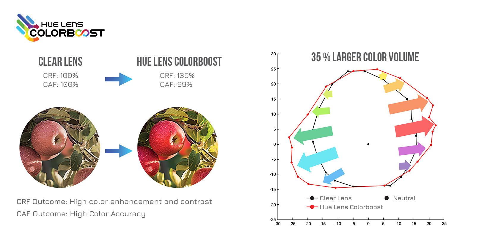
Fig. 3: A 35% larger color volume, visualized: more colors resolved, with accuracy held nearly level. Picture: Colorboost
Sources: Age-related color vision abnormalities: Schneck, M.E., et al. (2014), Optometry and Vision Science 91(1):60, DOI 10.1097/OPX.0000000000000114 (Smith-Kettlewell Eye Research Institute); contributing factors summarized by the California Health Report,
https://www.calhealthreport.org/2014/02/24/difficulty-seeing-some-colors-becomes-more-common-as-people-age/; lead author’s statement on blue-yellow defects: ESCRS EuroTimes, https://www.escrs.org/eurotimes/colour-vision; share of blue-yellow failures: Healio, https://cm.healio.com/news/optometry/20140224/certain-color-deficits-found-to-be-common-among-with-age. Gradual decline in normal aging: Barbur, J.L. & Rodriguez-Carmona, M. (2016), “Color vision changes in normal aging,” in Elliott, A.J., Fairchild, M.D. & Franklin, A. (Eds.), Handbook of Color Psychology, Cambridge University Press, pp. 180-196 (accepted manuscript), https://openaccess.city.ac.uk/id/eprint/12513/. Lens transmittance and age: Boettner, E.A. & Wolter, J.R. (1962), “Transmission of the ocular media,” Investigative Ophthalmology 1(6):776-783, https://ec.europa.eu/health/scientific_committees/scheer/docs/sunbeds_co240n_en.pdf. S-cone share of the cone population: Weinrich, T.W., et al. (2017), “No evidence for loss of short-wavelength sensitive cone photoreceptors in normal ageing of the primate retina,” Scientific Reports 7:46346, https://doi.org/10.1038/srep46346. S-cone pathway changes with age: “Aging of human short-wave cone pathways,” UC Davis, https://escholarship.org/uc/item/03×60251; “Effect of age on short-wavelength sensitive cone electroretinogram and long- and middle-wavelength sensitive cone electroretinogram,” https://pure.fujita-hu.ac.jp/en/publications/effect-of-age-on-short-wavelength-sensitive-cone-electroretinogra/. Peripheral resolution decline (~14% per decade after the fifth decade, for both blue-yellow and achromatic patterns): Zlatkova, M., Coulter, E. & Anderson, R. (2003), “Short-wavelength acuity: blue-yellow and achromatic resolution loss with age,” Vision Research, https://pure.ulster.ac.uk/en/publications/short-wavelength-acuity-blue-yellow-and-achromatic-resolution-los-3/. Debate and compensation: “The Verriest Lecture: Short-wave-sensitive cone pathways across the life span,” Journal of the Optical Society of America A 33(3):A104-22 (2016), https://pubmed.ncbi.nlm.nih.gov/26974914/; “Colour Constancy Across the Life Span: Evidence for Compensatory Mechanisms,” PLoS ONE (2013), https://doi.org/10.1371/journal.pone.0063921. CRF definition: Colorboost brand materials.